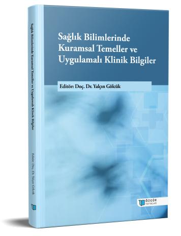
Bedside Percutaneous Tracheostomy in the Intensive Care Unit : Basic Principles and Current Developments
Chapter from the book:
Gölcük,
Y.
(ed.)
2025.
Theoretical Foundations and Applied Clinical Knowledge in the Health Sciences.
Synopsis
Percutaneous dilatational tracheostomy (PDT) is a minimally invasive procedure performed at the bedside in intensive care units. It is preferred in cases of prolonged mechanical ventilation or upper airway obstruction. Compared with endotracheal intubation, PDT offers several advantages, including improved patient comfort, reduced need for sedation, more effective clearance of secretions, and easier oral care.
Prior to the procedure, anatomical landmarks should be identified, and laboratory parameters (especially platelet count and coagulation tests) should be evaluated. Ultrasound can assist in identifying vascular structures and the puncture site. A team of at least three experienced physicians is recommended for the procedure. Performing the procedure under bronchoscopic guidance reduces complications. The tracheostomy tube should be selected according to the patient’s characteristics, and different sizes should be readily available.
Complications may occur in both the early and late phases. Early complications include hemorrhage, pneumothorax, pneumomediastinum, and malposition of the tube. Late complications include tracheal stenosis, fistula, tracheomalacia, and tube displacement. The most common complication is bleeding; therefore, careful evaluation of vascular anomalies is crucial.
In conclusion, PDT is a safe, cost-effective, and efficient method. Appropriate patient selection, an experienced team, and the use of ultrasound and bronchoscopy guidance can minimize complications.

